Home » Specialized Care » Orthopaedics » Diagnostic Imaging
Based on the clinical examination, we select the most suitable imaging techniques to confirm the suspected diagnosis and map further details.
X-rays provide clear and detailed insight into bone structures. We have both mobile wireless X-ray equipment and a fixed system available.
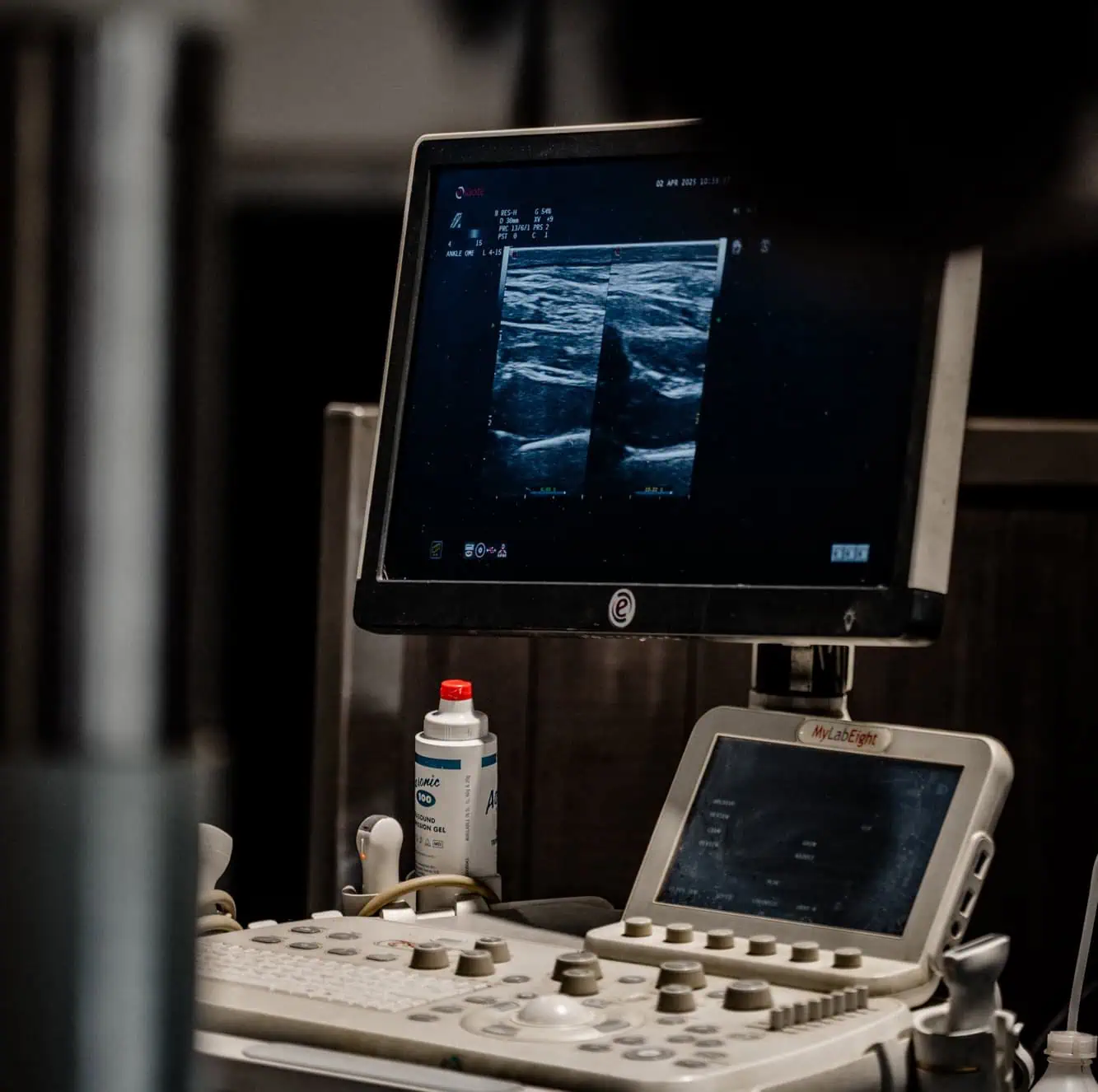
Ultrasound allows precise visualization of tendons, ligaments, muscles, and joint structures. Additionally, we use ultrasound for accurate, ultrasound-guided injections.
A CT scan provides a detailed, three-dimensional view of the horse’s bones and soft tissues. This advanced technique is particularly suited for examining complex regions such as the neck, head, and limbs (including shoulder and pelvis). CT helps us detect fractures, bone abnormalities, joint problems, and cartilage or tendon injuries that may go unnoticed with conventional techniques.
MRI is an advanced three-dimensional examination of bones and soft tissues, including ligaments, tendons, and joints. With standing MRI, the horse’s lower limbs can be imaged from hoof to carpus or hock. This makes it especially suitable for the early detection of subtle injuries that remain invisible with other techniques.
UTC provides detailed information about the structure and quality of tendon tissue. It is an indispensable tool for diagnosing chronic tendon problems, monitoring recovery, and identifying weak spots at an early stage.
Trust in the excellent care provided by Via Nova’s specialists.