Home » Specialized Care » Advanced imaging
MRI is an advanced three-dimensional examination of bones and soft tissues, including ligaments, tendons, and joints. With standing MRI, the horse’s lower limbs can be imaged from hoof to carpus or hock. This makes it especially suitable for the early detection of subtle injuries that remain invisible with other techniques.
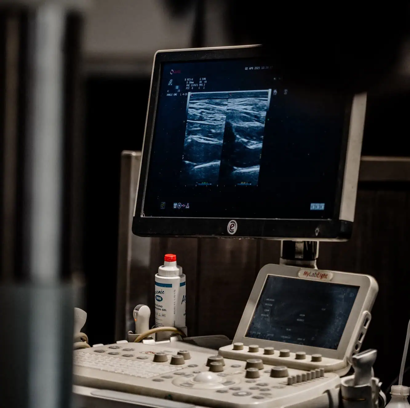
Interpreting radiographic, ultrasonographic, and other diagnostic images requires specialist knowledge and experience. At Via Nova, a board-certified specialist reviews your imaging for a reliable analysis.
For a second opinion, you are also welcome to consult us. Our specialist carefully assesses your images and medical records and provides clear and expert advice.
Trust in the excellent care provided by Via Nova’s specialists.